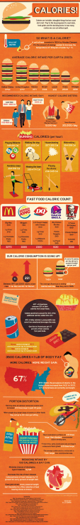
Heart Anatomy
The heart is one of the most important organs in the human body. It is essentially a 2-sided pump that runs on electricity to push blood around your body. Heart anatomy consists from these main parts and their functions:
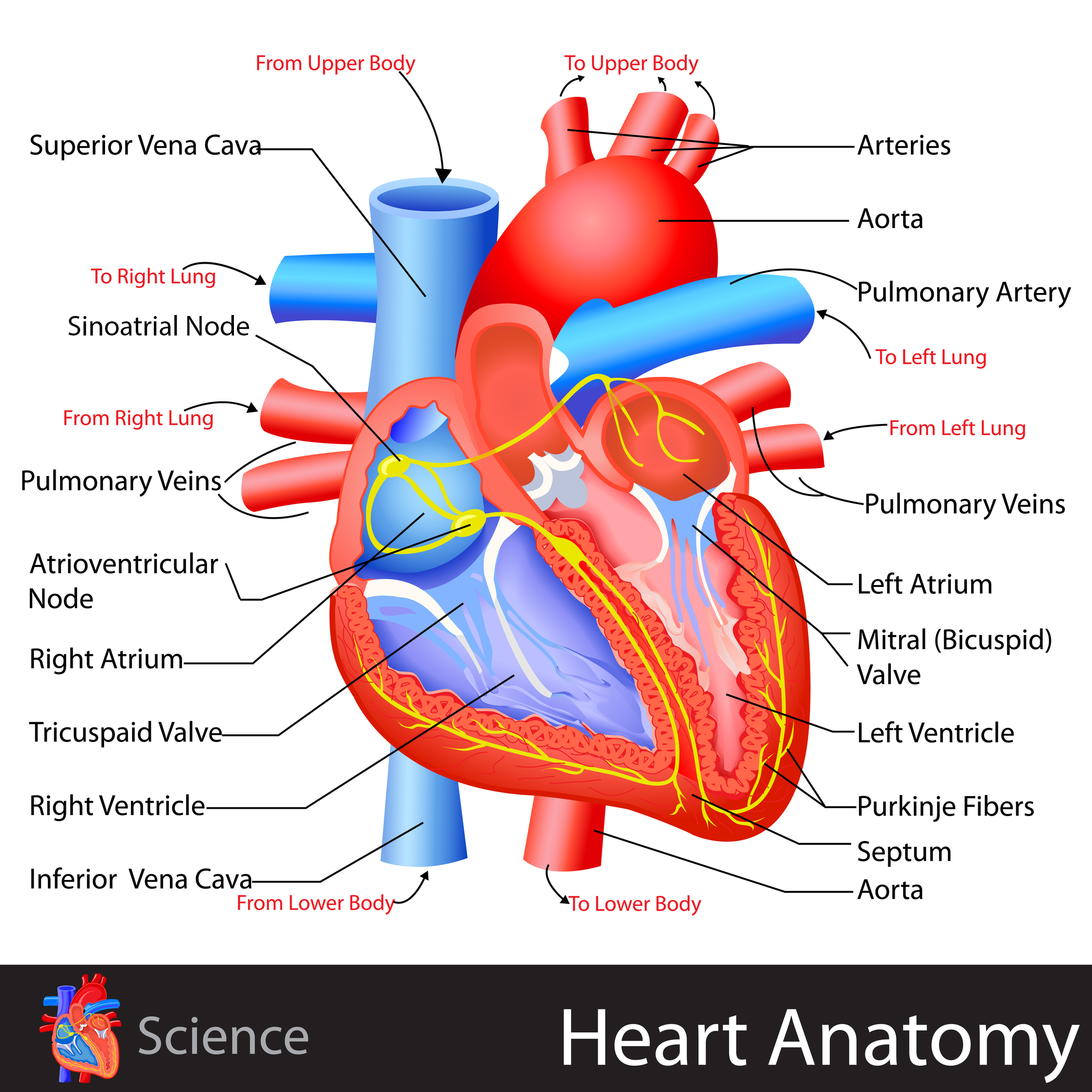
Blood Vessels
Veins carry blood to the heart, while arteries carry blood away from the heart.
Superior Vena Cava – main vein carrying deoxygenated blood into the right side of the heart.
Pulmonary Arteries – carry deoxygenated blood from the right side of the heart to the lungs where oxygen is added.
Pulmonary Veins – carry oxygen-rich blood back to the left side of the heart where it can be pumped to the rest of the body.
Aorta – main artery carrying oxygen-rich blood from the left side of the heart to the rest of the head and body.
Chambers and Separations
The heart has 2 sides separated by the septum, and each side consists of an atrium and a ventricle separated by valves.
Right Atrium – fills up with deoxygenated blood from the body.
Tricuspid Valve – 3 sided valve that controls blood flow between right atrium and ventricle.
Right Ventricle – pumps blood out to the lungs to be oxygenated.
Left Atrium – fills up with oxygen-rich blood from the lungs.
Mitral (Bicuspid) Valve – 2-sided valve that controls blood flow from the left atrium into the left ventricle
Left Ventricle – pumps blood out to the body
Electrical System
Sinoatrial Node – bundle of tissue that creates the electrical impulses for the heart contraction.
Atrioventricular Node – bundle of tissues which controls the flow of electrical impulses to the ventricles.
Purkinje Fibres – nerve fibers that carry electrical signals into ventricle walls and signal contractions.
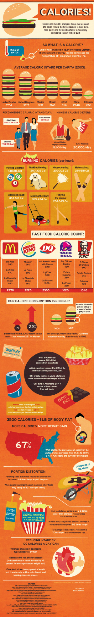
You are done with the heart anatomy? Check out our interesting article about the tongue!